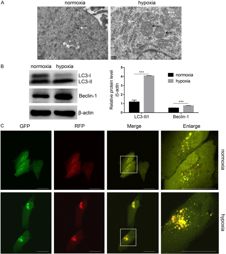
Figure 6.

Hypoxia pretreatment promotes cell autophagy. A. Autophagosomes were observed under transmission electron microscopy (TEM). B. Autophagy-related markers LC3 and Beclin1 were analyzed by western blot with β-actin normalization. C. USCs were transfected with ptfLC3 and treated as above described. Autophagic flux was dynamically observed by laser scanning confocal microscope. ***P<0.001, Student’s t-test. Scale bar =200 μm. (White arrow indicates autophagosome, black arrow indicates autophagic lysosome).
