Figure 1.

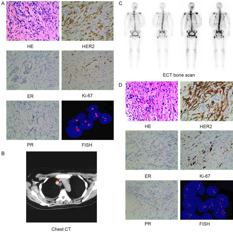
Histopathologic, molecular and imaging characterization identified the patient as HER2-positive bilateral breast cancer with distant metastases. A. HE staining showed infiltrating ductal carcinoma (×400). Immunohistochemical staining (×400) showed the tumor was negative for ER (-) and PR (-), but positive for HER2 (2+~3+) and Ki-67 (+, 20%). FISH assay revealed HER2 gene amplification of infiltrating ductal carcinoma from right breast of the patient. B. Tumor burden of metastatic mediastinal lymph nodes (arrow) as evidenced by chest CT scan. C. Multiple bone metastases as evidenced by ECT bone scanning. D. HE staining showed infiltrating ductal carcinoma (×400). Immunohistochemical staining (×400) showed the tumor was negative for ER (-), PR (-), positive for HER2 (3+), and Ki-67 (+, 50%). FISH assay revealed HER2 gene amplification of infiltrating ductal carcinoma from left breast of the patient. Abbreviations: CT, computed tomography; ECT, emission computed tomography; ER, estrogen receptor; FISH, Fluorescent in situ hybridization; HE, hematoxylin & eosin; HER2, human epidermal growth factor receptor 2; PR, progesterone receptor.
