Figure 2.

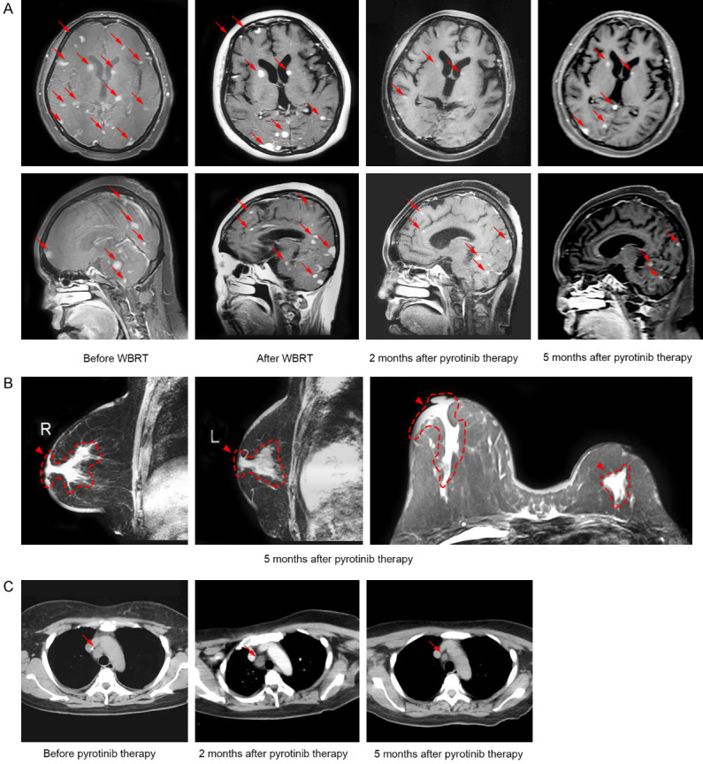
Imaging characterization revealed dynamic changes of the primary and metastatic lesions during the treatment of pyrotinib. A. Cranial MRI revealed continuous shrinkage of brain metastases (arrows) after WBRT and pyrotinib therapy. B. Breast MRI revealed reductions in primary breast lesions (arrowheads) 5 months after pyrotinib therapy. C. Chest CT scan revealed a significant reduction in lesion of mediastinal lymph node (arrows) 5 months after pyrotinib therapy. Abbreviations: CT, computed tomography; MRI, magnetic resonance imaging; WBRT, whole brain radiotherapy.
