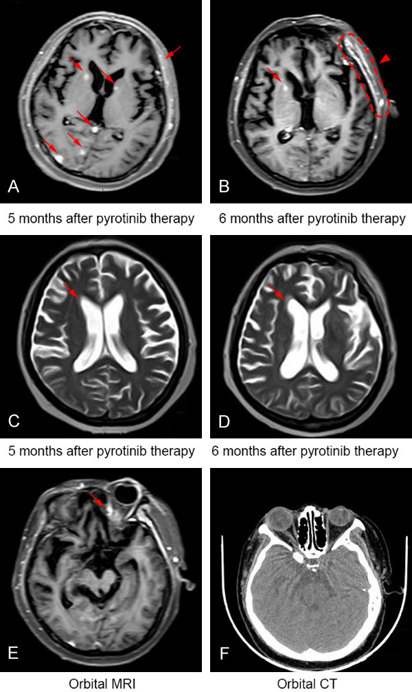
Figure 3.

The patient showed tumor response discordance between intracranial parenchymal and meningeal metastatic lesions when receiving further treatment of pyrotinib. A and B. While a sustained relief of intracranial parenchymal lesions (arrows) can be observed by pyrotinib therapy for 6 months, a band-like high signal (arrowhead) in left frontotemporal dura was revealed by cranial MRI. C and D. Obtuse horn of the ventricle (as indicted by arrow) was shown by cranial MRI 6 months after pyrotinib therapy. E. A suspected high signal (arrow) in left orbital apex was shown by cranial MRI. F. Lateral orbital CT scan confirmed no metastasis of the tissue. Abbreviations: CT, computed tomography; MRI, magnetic resonance imaging.
