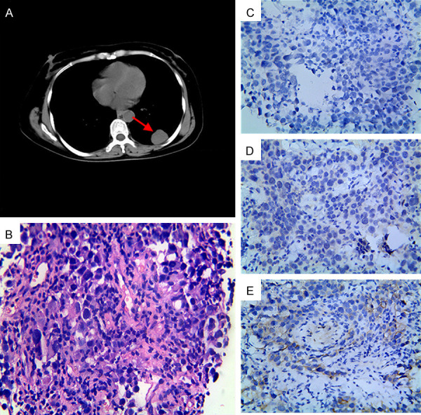
Figure 5.

Imaging and histopathologic characterization revealed a left lung metastasis with a triple-negative pathological feature. A. CT scan showed a lesion in the left lower lung, as indicated by the arrowhead; B. HE staining indicated the lesion as metastatic infiltrating carcinoma (400×); C-E. Immunohistochemical staining showed the metastatic lesion was negative for ER, PR, and HER2 (400×). Abbreviations: BC, breast cancer; CT, computed tomography; ER, estrogen receptor; HE, hematoxylin & eosin; HER2, human epidermal growth factor receptor 2; PR, progesterone receptor.
