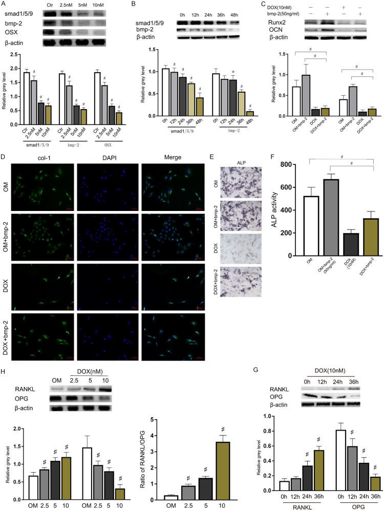
Figure 3.
DOX restrained the bmp-2/smad signalling pathway of osteogenesis differentiation in BMSCs. A. BMSCs cultured with OM (control), DOX (2.5, 5, 10 nM) were harvested on days 2. The protein expression levels of smad1/5/9, bmp-2, and OSX were assessed by WB and quantified *P<0.05 and #P<0.01 vs the control group. B. The protein expression levels of smad1/5/9 and bmp-2 were quantified *P<0.05 and #P<0.01 vs the control group. C. BMSCs cultured with OM (control), with or without DOX (10 nM) or bmp-2 (50 ng/ml) were harvested on 48 h, the protein expression of Runx-2 and OCN were quantified. *P<0.05 and #P<0.01 vs the control group. D. Immunofluorescence detection of col-1 translocation in cultured. BMSCs were treated with OM (control), with or without DOX (10 nM) or bmp-2 (50 ng/ml). Col-1 expressed in both the cytoplasm and nucleus, as well as the fluorescent density and intensity was increased dose dependently for 5 days. The nuclei were stained with DAPI and were shown as blue fluorescence. Scale bar = 100 µm. E, F. BMSCs cultured with OM (control), with or without DOX (10 nM) or bmp-2 (50 ng/ml) were harvested on 7 days, ALP staining/activity was assessed. G. BMSCs treated with OM (control), DOX (10 nM) were harvested on 0 h, 12 h, 24 h, 36 h. The protein expression levels of RANKL and OPG were assessed by WB and quantified *P<0.05 and #P<0.01 vs the control group. H. BMSCs treated with OM (control), DOX (2.5, 5, 10 nM) were harvested on 48 h. The protein expression levels of RANKL and OPG were assessed by WB and quantified *P<0.05 and #P<0.01 vs the control group.

