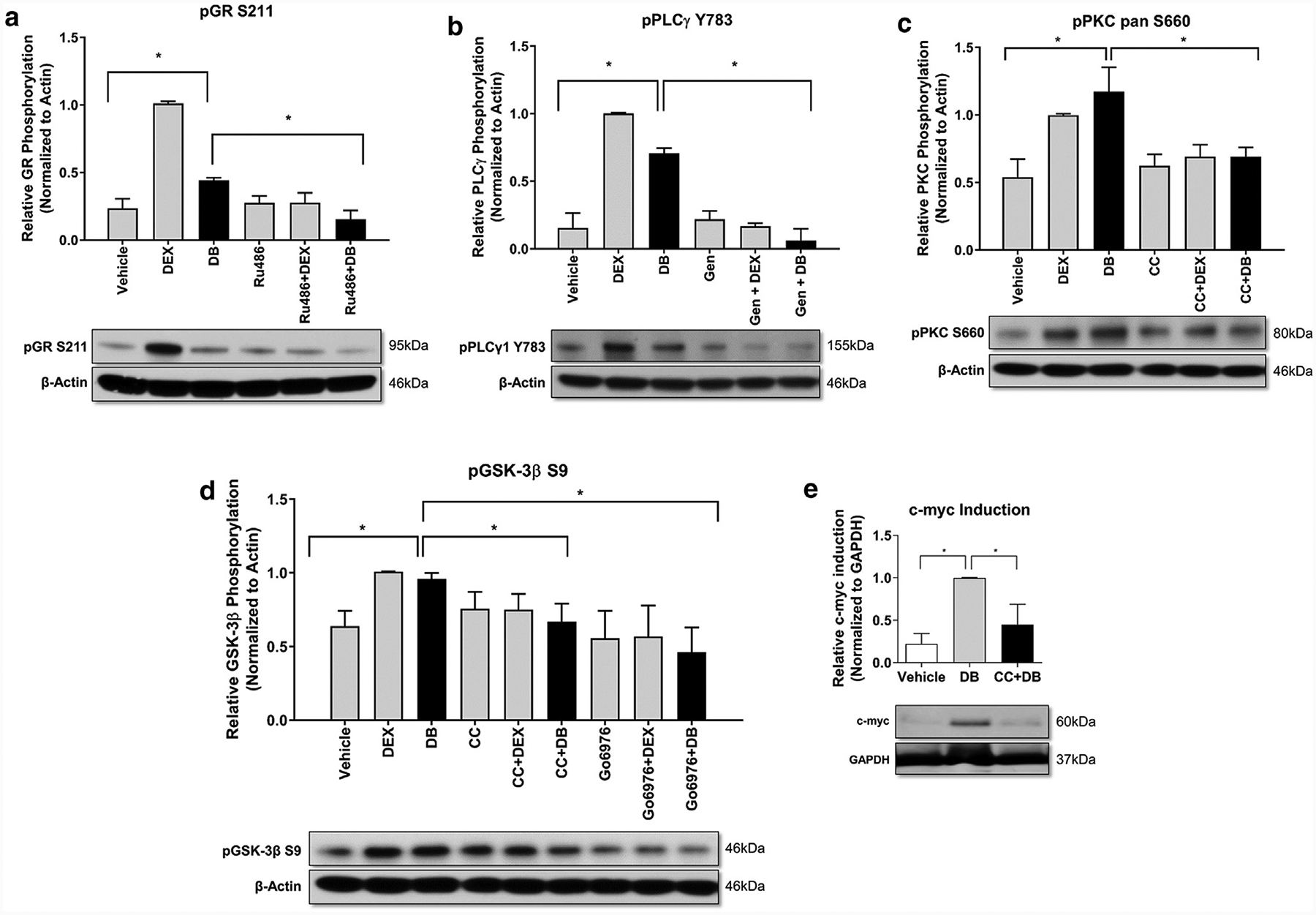
Figure 2. mbGR stimulation activates PLC/PKC signaling cascade and induces phosphorylation and subsequent inactivation of GSK-3β resulting in c-myc induction.

Cells were stimulated with vehicle (DMSO), 1 μM Dex, or 100 nM Dex-BSA (DB) for 30 minutes upon treatment with GR antagonist Ru486 (1 μM), PLCγ inhibitor, genistein (50 μM) or PKC inhibitors CC (200 nM) or Go6976 (4μM). Phosphorylation of GR (a), PLCγ (b), PKC (c), and GSK-3β (d) were assessed by western blot. (e) Cells were stimulated with vehicle (DMSO) or 100 nM Dex-BSA for 4 hours upon treatment with 200 nM CC, and induction of c-myc was assessed by western blotting. All quantifications were performed using ImageJ with error bars corresponding to standard deviation from n = 3. CC, calphostin C; Dex, dexamethasone; Dex-BSA, BSA-conjugated dexamethasone (DB); GR, glucocorticoid receptor; GSK-3β, glycogen synthase kinase 3 beta; mbGR, membranous glucocorticoid receptor; PKC, protein kinase C; PLC, phospholipase C. *P ≤ 0.05 (Student t test).
