
Figure 5. Selective targeting of mbGR by Dex-BSA impedes epithelialization and can be ameliorated by inhibiting PKC in an ex vivo model of wound closure.
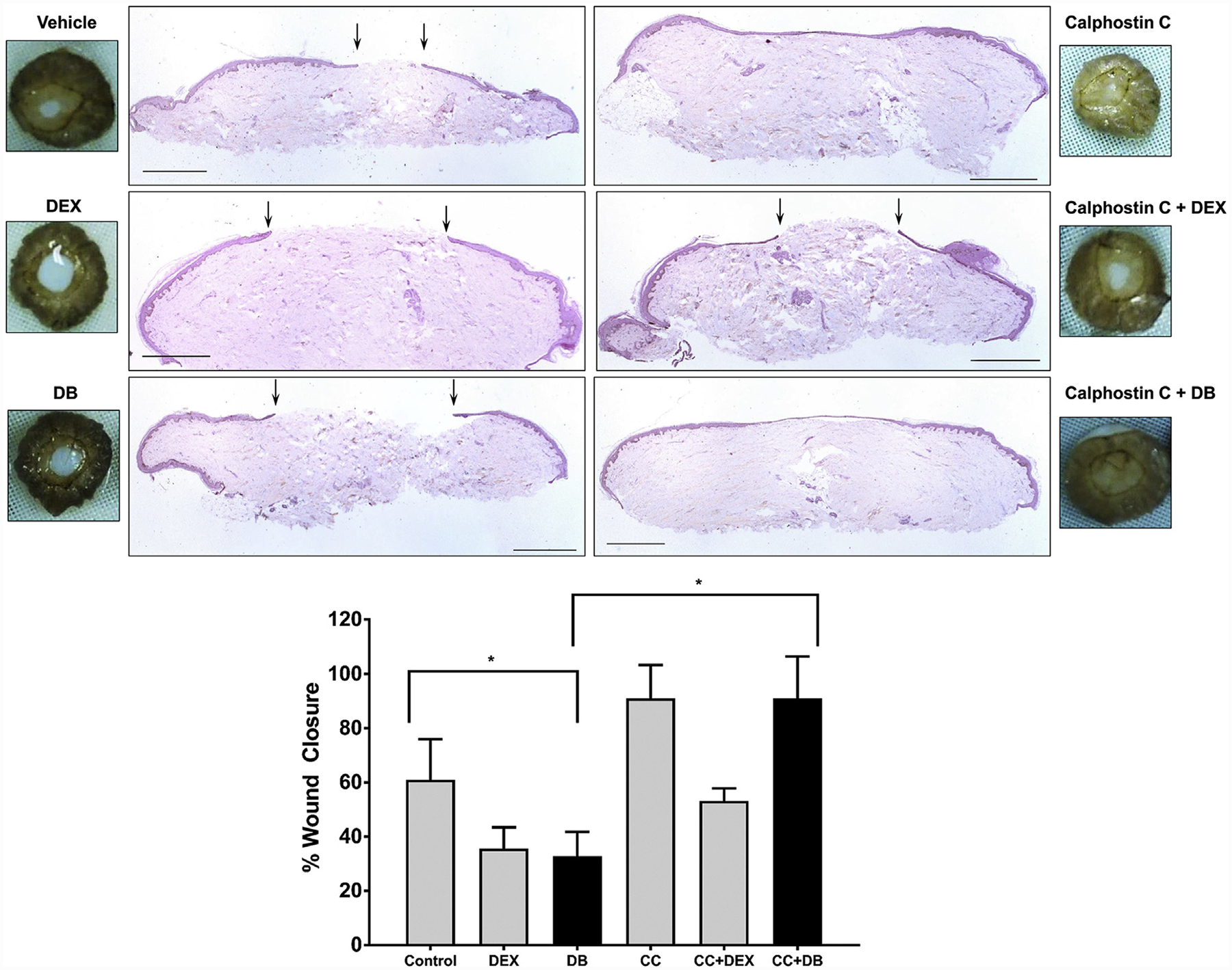
Normal human skin was wounded using a 3-mm biopsy punch and maintained at the air-liquid interface in presence or absence of 1 μM Dex, 100 nM Dex-BSA (DB), or PKC inhibitor CC (0.2 μM). Wound healing was assessed at day 4 after wounding, a time when exponential epithelialization occurs. Wound closure was quantified by histology using ImageJ software. Gross photos show visual signs of closure and correspond to the histology assessments with black arrows pointing to epithelial tongue location at day 4. Error bars correspond to standard deviation from n = 4. *P ≤ 0.05 (Student t test). Scale bar 1 = mm. Dex, dexamethasone; Dex-BSA, BSA-conjugated dexamethasone (DB); mbGR, membranous glucocorticoid receptor; PKC, protein kinase C.