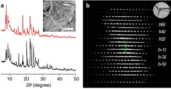
Figure 1.

a) PXRD patterns of the as‐made (black) and calcined (red) forms of pure‐silica PST‐24. Inset: SEM image of its as‐made form (scale bar: 1 μm). b) 3D reciprocal lattice of calcined, pure‐silica PST‐24 reconstructed from cRED data viewed along the c‐axis. While the diffraction spots with k=2n are sharp, those with k=2n+1 are shown as diffuse streaks. Inset: the crystal where the cRED was collected.
