
Figure 1.
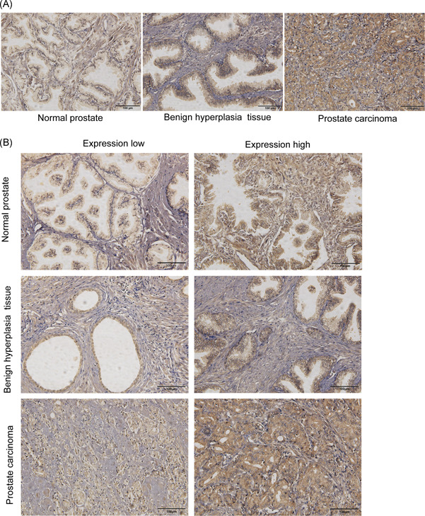
Representative images and scoring of immunohistochemical staining of β2‐AR. A, Immunohistochemistry images of β2‐AR expression in the tissues of TMA sections. Scale bar, 100 μm. B, Examples of scoring low and high immunohistochemical stains for β2‐AR are shown. β2‐AR, β2‐adrenergic receptor [Color figure can be viewed at wileyonlinelibrary.com]