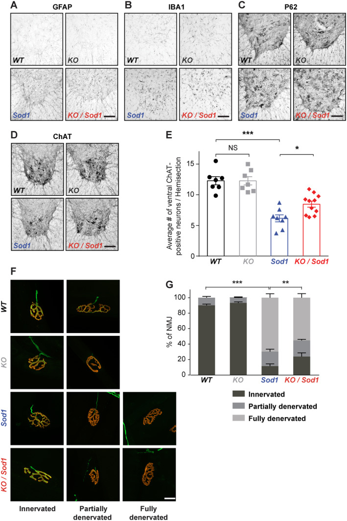
FIGURE 4.

Absence of corticospinal neurons (CSN) partly prevents degeneration of the motoneuron (MN) cell bodies and neuromuscular junction (NMJ) dismantlement. (A–D) Representative immunostaining images of the ventral horn of the lumbar spinal cord from end‐stage Sod1 and KO/Sod1 mice and their age‐matched WT and KO littermates labeled with GFAP (A), IBA1 (B), P62 (C), and choline acetyltransferase (ChAT; D). (E) Bar graph representing the average number of ventral ChAT+ neurons per lumbar spinal cord hemi‐section; 1‐way ANOVA; n = 6 WT, n = 6 KO, n = 8 Sod1, and n = 10 KO/Sod1. (F) Representative maximum‐intensity projection images of z‐stacks of typically innervated, partly or fully denervated NMJs from end‐stage Sod1 and KO/Sod1 mice and their age‐matched WT and KO littermates. (G) Bar graph representing the average proportions of innervated (dark gray), partly denervated (medium gray) and fully denervated (light gray) NMJs for each genotype; 2‐way ANOVA followed by Tukey multiple comparisons test; n = 6 animals per genotype. **p < 0.05, **p < 0.01, ***p < 0.001; NS, nonsignificant. Scale bars: 100μm in A–D; 20μm in F. [Color figure can be viewed at www.annalsofneurology.org]
