
Figure 6.
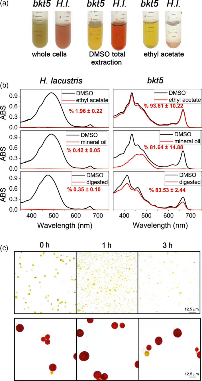
Extractability and bio‐accessibility of astaxanthin from Haematococcus lacustris and Chlamydomonas reinhardtii cells. (a) Aplanospore cysts of H. lacustris in ‘red phase’ and C. reinhardtii bkt5 in liquid medium (left) were compared using different pigment extraction methods. DMSO is used as a control from total extraction of pigments from both hosts (centre) and ethyl acetate (right) or mineral oil were used. (b) Absorption spectra of pigments extracted from H. lacustris in red phase and bkt5 with different solvents. Black traces are the absorption spectra of pigments obtained upon extraction in DMSO (positive control: maximum extraction); red traces show the pigments extracted with different methods. Spectra are normalized to the maximum absorption of the DMSO control. Inserts indicate the percentage of carotenoids extracted calculated from fitting analysis of the absorption spectra as described in Croce et al. (2000). Data are expressed as mean ± SD (n = 3). (c) Images of cells of bkt5 and H. lacustris before (0 h) and after simulated gastro‐intestinal digestion (1 or 3 h).