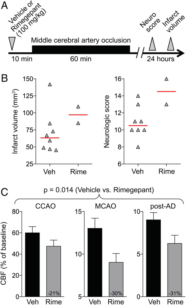
FIGURE 3.

Rimegepant worsens the outcomes after 60‐minute focal cerebral ischemia. (A) Experimental timeline. (B) Infarct volume (mm3) and neurologic deficit scores. Data from individual animals are shown along with their group median (red lines). Mean ages were 2.2 ± 0.2 and 2.3 ± 0.3 months in vehicle (Veh) and rimegepant (Rime; 100mg/kg) groups, respectively (all male, wild type; mean ± standard deviation). (C) Residual cerebral blood flow (CBF; % of baseline) during middle cerebral artery occlusion (MCAO; p = 0.014, 2‐way repeated measures analysis of variance). Numbers within the gray bars represent the relative difference between treatment arms calculated as (CBFOlce/CBFVeh) − 1. One mouse each in the vehicle and rimegepant arms were excluded from analyses based on predetermined criteria (see Materials and Methods). Six mice died prior to outcome assessments in the rimegepant arm. AD = anoxic depolarization; CCAO = common carotid artery occlusion. [Color figure can be viewed at www.annalsofneurology.org]
