Figure 1.

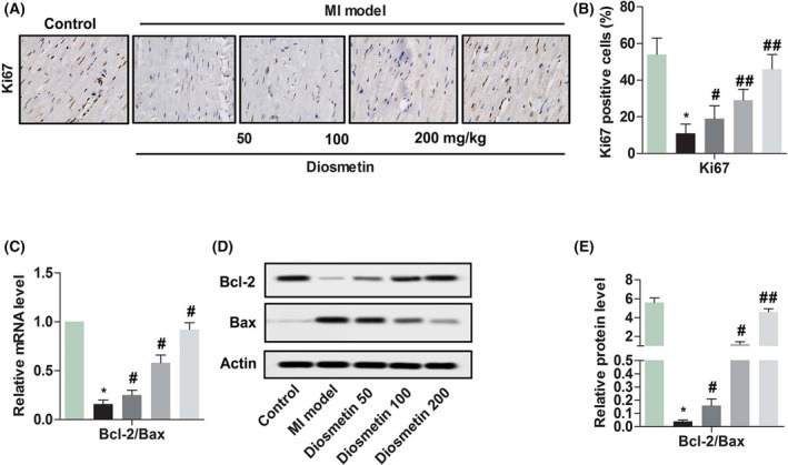
Diosmetin enhances the proliferation marker proteins of Ki67 and expression of the ratio of Bcl‐2/Bax in MI model rats. A, Comparison of Ki67 expression proteins in myocardial cells detected by immunohistochemical staining from Sham group, MI model group, low‐dose group (50 mg/kg diosmetin), medium‐dose group (100 mg/kg diosmetin) and high‐dose group (200 mg/kg diosmetin). Magnification 200×. B, Semi‐quantitative analysis of the relative amounts of Ki67 in each group of neonatal rats. C, Relative mRNA level ratio of Bcl‐2/Bax detected by RT‐qPCR in each group of neonatal rats. D, A representative result for western blot analysis Bcl‐2 and Bax. E, Semi‐quantitative analysis of the relative amounts’ ratio of Bcl‐2/Bax in each group of neonatal rats. The results were presented as mean ± SD and represent three individual experiments. (*P < .05 vs sham group, #P < .05 vs MI model group, ##P < .01 vs MI model group)  , Control;
  , MI model;
  , Diosmetin 50;
  , Diosmetin 100;
  , Diosmetin 200;
  , Control;
  , MI model;
  , Diosmetin 50;
  , Diosmetin 100;
  , Diosmetin 200;
  , Control;
  , MI model;
  , Diosmetin 50;
  , Diosmetin 100;
  , Diosmetin 200
