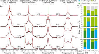
Figure 1.

A) Static 2H NMR spectra (black) of the SC lipid model mixture composed of Cer[NS]/LA/Chol at molar ratios of 1:1:0.45 (left two columns) and 1:1:1 (right two columns) at various temperatures. The first and third column display the 2H NMR spectra of the Cer[NS]‐sph‐d 28 (deuterated sphingosine‐d 28). Columns 2 and 4 show the NMR spectra of the Cer[NS]‐acyl‐d 47 (perdeuterated lignoceroyl‐d 47). Simulated spectra are shown in red. The multilamellar lipid preparations contained 50 wt % buffer (0.1 m MES, 0.1 m NaCl, 5 mm EDTA, pH 5.4). B) Relative proportions of the individual phases observed in the mixtures.
