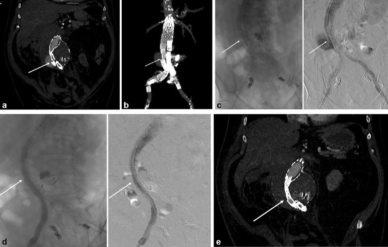
Fig. 1.

Late type III endoleak due to iliac component separation in a Cook Zenith placed 10 years prior to presentation. The patient had been lost to follow-up for several years and presented with a contained rupture. ( a ) Reformatted CT angiography at presentation, demonstrating separation between the main body of the graft and the right iliac limb. Arrow denotes the separation between the main body and the right iliac limb. ( b ) Three-dimensional reconstruction of presenting CT angiography. Arrow again denotes the separation between the main body and the right iliac limb. ( c ) Intraoperative angiography demonstrating extravasation. Arrow denotes the separation between the main body and the right iliac limb. ( d ) intraoperative angiography after placement of a bridging iliac limb with appropriate covering of the prior graft component separation (arrow). ( e ) CT angiography performed prior to discharge demonstrating successful repair, with arrow denoting position of the bridging iliac limb.
