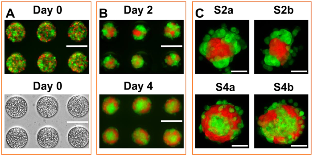
Figure 1: Different architectures of co-culture spheroids formed within an array microwell platform.

Co-culture tumor spheroids were formed by mixing two cell types in an array microwell. A. Fluorescence (top panel) and bright field (bottom panel) micrographs of 1:1 ratio of two cell types in microwells at day 0 using an epi-fluorescence microscope. B. Florescence image of co-culture spheroids at day 2 and day 4. Images were taken at the mid - Z plane of the spheroids using an epi-fluorescence microscope. C. Close-up confocal images of four types of spheroid architecture observed at day 2 and day 4. Images were reconstructed as projection of Z planes of the entire spheroids. Spheroids on day 2 exhibited a morphology with metastatic MDA-MB-231 cells outside and nontumorigenic MCF-10A cells inside (S2a and S2b). Spheroids on day 4 presented a reversed morphology with MCF-10A cells surrounding MDA-MB-231 cells, with a few loosely MDA-MB-231 cells attaching the periphery (S4a and S4b). In S2a, and 4a, one cell type were enclosed by the other cell type. In S2b, 4b, one cell type only partially surrounded by the other cell type. Green cells were malignant breast tumor cell line (MDA MB-231) expressing green fluorescent protein; red cells were a non-tumorigenic breast epithelial cell line (MCF-10A) expressing dTomato. Scale bar: 200 μm in A and B and 50 μm in C.
