FIGURE 1.

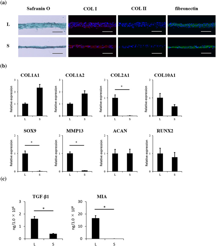
Properties of LC sheets and SY sheets. For each sheet, n = 6. (a) Histological analysis of LC sheets and SY sheets revealed negative staining for Safranin O. Immunohistochemical analysis revealed positive staining for COL I and fibronectin, but negative staining for COL II for both sheet types. Scale bar = 50 μm. (b) Gene expression profile of LC sheets and SY sheets. mRNA expression for COL2A1, SOX9, and MMP13 was significantly higher in LC sheets than in SY sheets (p < 0.05). (c) The concentrations of humoral factors secreted by LC sheets and SY sheets. The secretion of TGF‐β1 was 1.6 ± 0.2 ng/1.0 × 106 cells by LC sheets and 0.40 ± 0.02 ng/1.0 × 106 cells by SY sheets. The secretion of MIA by LC sheets was 16.5 ± 2.1 ng/1.0 × 106 cells, compared with undetectable levels secreted by SY sheets. The secretion of MIA and TGF‐β1 by LC sheets was significantly higher than that by SY sheets (p < 0.05). The results are presented as mean ± standard deviation. LC: layered chondrocyte; SY: synoviocyte; COL I: type I collagen; COL II: type II collagen; COL1A1: collagen, type I, alpha 1; COL1A2: collagen, type I, alpha 2; COL2A1: collagen, type II, alpha 1; COL10A1: collagen, type X, alpha 1; SOX9: SRY‐Box 9; MMP13: matrix metalloproteinase 13; ACAN: aggrecan; RUNX2: Runt‐related transcription factor 2; TGF‐β1: transforming growth factor‐β‐1; MIA: melanoma inhibitory activity; L: LC sheets; S: SY sheets [Colour figure can be viewed at wileyonlinelibrary.com]
