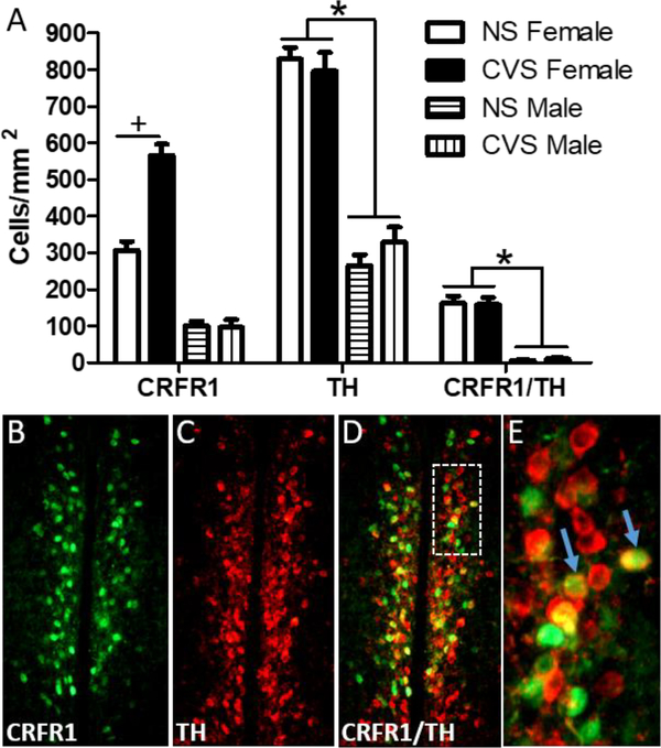
Figure 5. Co-localization of AVPV CRFR1-GFP with TH following CVS.
(A) The number of CRFR1/TH co-localized cells was unaffected in male and female mice following CVS. This indicates that the increase in CRFR1-GFP labeled cells in CVS females occurs in an independent set of neurons. (B-D) Representative images of CRFR1-GFP, TH, and a merged image from a CVS female mouse, respectively. White inset box (D) indicates the area further magnified in (E). Arrows show examples of co-labeled neurons. * indicates a greater number of CRFR1-GFP, TH, and co-labeled cells in females compared to males, p < .001. + indicates p < .001 (NS compared to CVS females).

