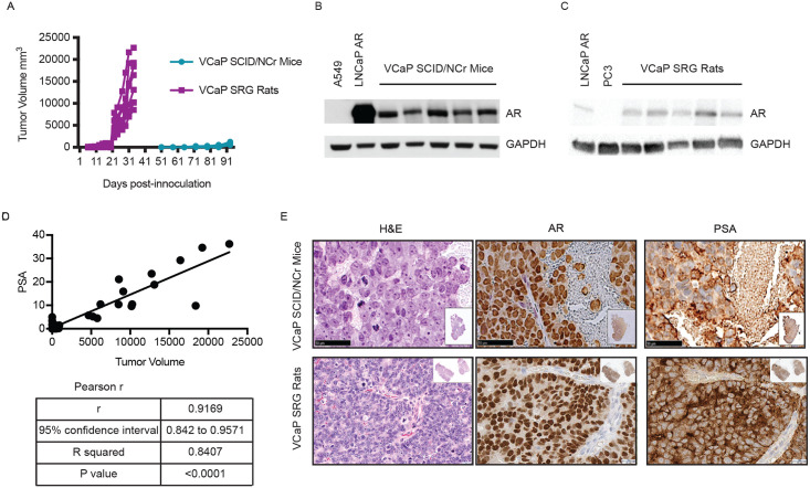
Fig 4. VCaP xenograft model in SCID/NCr mouse and SRG rat.
SCID/NCr mice and SRG rats were inoculated with 5x10⁶ and 10x106 VCaP cells, respectively, subcutaneously in the hind flank. Tumor width and length were measured three times weekly to calculate volume. A) Tumor kinetics in the SRG rat vs. SCID/NCr mouse. Each line represents tumor growth in an individual SRG rat or SCID/NCr mouse. B) Western blotting for AR in tumor tissue from the SCID/NCr mice. C) Western blotting for AR in tumor tissue from the SRG rat. D) Compilation of PSA in the serum of SRG rat inoculated with VCaP cells correlates with tumor volume. E) H&E staining and IHC staining for AR and PSA in VCaP tumor tissue from SCID/NCr mice and SRG rat.

