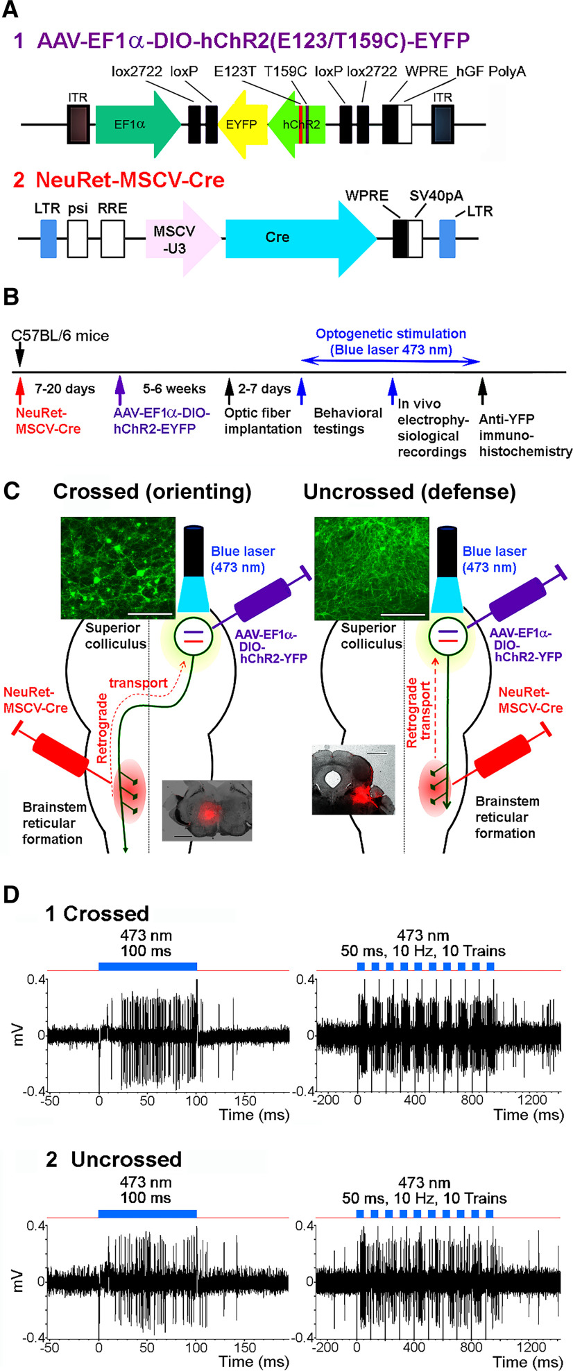
Figure 1.
Pathway-selective optogenetic activation of SC output neurons. A, Viral vector constructions. B, Experimental protocols. C, Schematic diagram for the double injection of the viral vectors into the brainstem and SC, and the interaction of NeuRet-MSCV-Cre and AAV-EF1α-DIO-hChR2(E123T/T159C)-EYFP in the double infected SC neurons. Upper insets in left and right panels, Photomicrographs of the somata of the crossed (orienting) and uncrossed (defense) SC-brainstem pathway neurons, respectively. Lower insets in left and right panels, Injection sites of the NeuRet-MSCV-Cre, indicated by a mock injection of Fluoro-Ruby into the brainstem reticular formation at medial PMRF and CnF, respectively. Scale bar in the upper insets = 100 μm. Scale bar in the lower insets = 1 mm. D, Responses of a mouse SC output neuron to blue laser stimulation (blue line) with a 500-μm diameter fiber. (1) Responses of the crossed (orienting) pathway neurons. (2) Responses of uncrossed (defense) pathway neurons. Figure Contributions: Thongchai Sooksawate, Kenta Kobayashi, and Kaoru Isa performed the experiments. Thongchai Sooksawate analyzed the data and prepared the figure.

