
Figure 6. Oligodendrocyte depletion and demyelination following cuprizone administration impairs multi-unit recording activity.
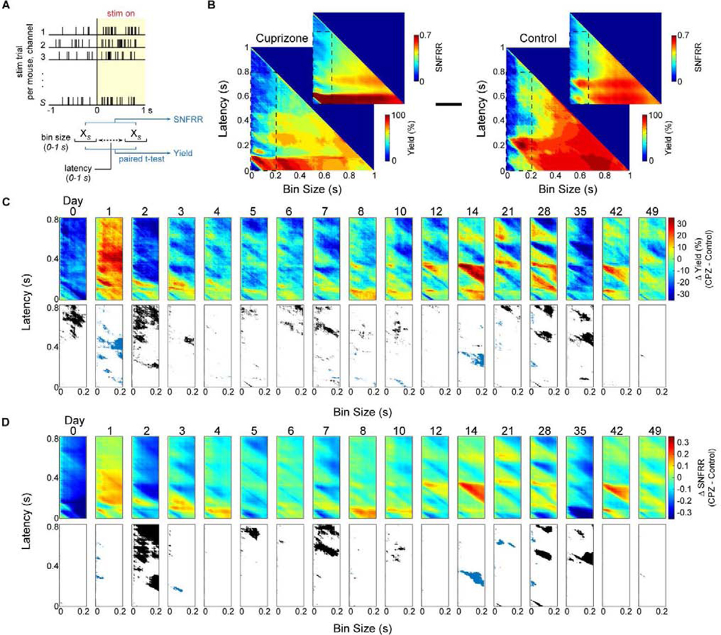
(a) Diagram demonstrating how dynamic changes in multi-unit yield and SNFRR during evoked electrophysiological recordings were measured. Spike counts, XS, were collected within a temporal bin size and latency ranging from 0 to 1 s following stimulus presentation and compared to counts within the same bin size before stimulus presentation using a paired t-test. MU yield was calculated as the percentage of electrode channels (out of 16 sites) that had a significantly different spike count for a given bin size and latency after stimulus presentation compared with the same bin size before stimulus presentation. MU SNFRR was calculated as the difference in average spike counts relative to the standard deviation in spike counts before and after stimulus presentation. (b) Representative plots of MU yield and SNFRR taken at varying bin sizes and latency between 0 and 1 s at 1 ms increments in cuprizone-treated and control mice. MU yield and SNFRR plots within a window of 200 ms bin size and 800 ms latency (inset) were averaged within each group and control data was subtracted from cuprizone mice to produce (c) and (d). (c-d) Difference in MU yield or SNFRR between cuprizone-treated and control mice over time (top panel). A paired t-test (p < 0.05) was performed at each bin size and latency between cuprizone-treated and control mice. Significant differences where yield or SNFRR was greater in control mice are reported in black, where yield or SNFRR was greater in cuprizone mice are reported in blue, and no significant difference between the two groups are reported in white (bottom panel).