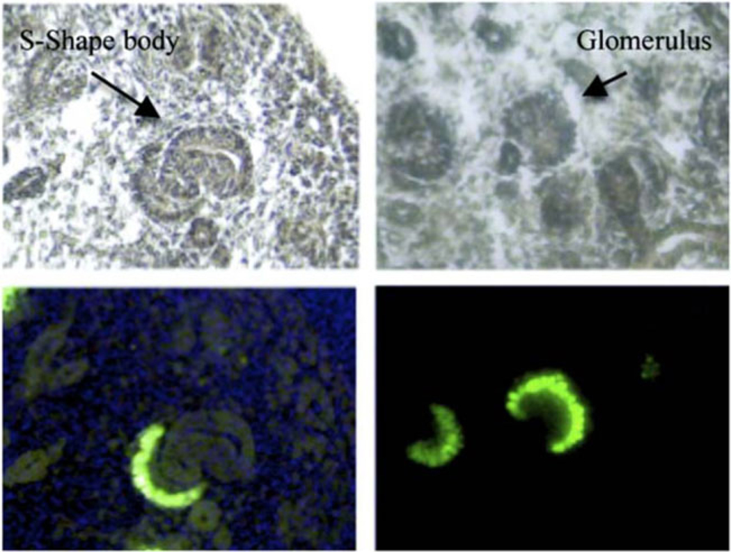
Fig. 1.
MafB-GFP mice show green fluorescent protein (GFP) expression restricted to podocytes. Top panels show cryostat sections with S-shaped body (left) and forming glomerulus (right) marked by arrows. Bottom panels show that GFP fluorescence is restricted to developing podocytes at E15.5. (Originally published in [20]

