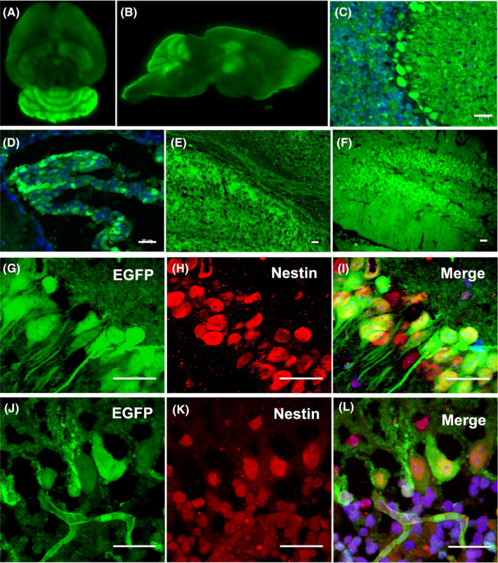
Figure 2.

Direct observation of Foxn1nu.B6‐Tg(CAG‐EGFP) mice under fluorescence microscope (480 nm): (A) horizontal position of brain; (B) sagittal position of brain; (C) cerebellar cortex; (D) lateral ventricle choroid plexus; (E) corpus callosum; and (F) hippocampus. G‐L, Immunofluorescence staining of EGFP and Nestin in hippocampal dentate gyrus (upper panels) and cerebellar Purkinje cells (lower panels). Confocal laser scanning microscopy revealed that a few cells were EGFP/Nestin double positive. Scale bar = 50 µm
