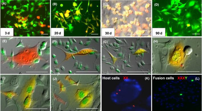
Figure 7.

Observation of SU3/RFP co–cultured with peritoneal lavage cells under fluorescence microscope and Live Cell Imaging System in vitro. A‐D, Yellow co–localized cells at different time points of red fluorescence protein (RFP) enhanced green fluorescent protein(EGFP) merged images, wherein “yellow cells” in image C are monoclonal cells from image B, and image D is long‐term subcultured cells of image C, in which the co–localized yellow cells were gradually rare. E‐G, An RFP cell phagocytized an EGFP cell, before splitting into two co–localized “yellow cells.” H‐J, A binuclear yellow cell divided into two cells with green nuclei and yellow cytoplasm. K‐L, Sex chromosome‐specific FISH assay detected the karyotype of “yellow cells.” Red fluorescence was labeled in chromosome X probe; chromosome Y probe was labeled with green fluorescence. SU3 cell was from a male patient. FISH assay showed that karyotype of SU3 cells was XY, which has been published in a previous report; tumor‐bearing mice were all female, FISH assayed showed karyotype of host cells was XX (K); The “yellow cells” were identified in XXXY (L). Scale bar = 50 µm
