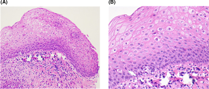
FIGURE 3.

Individual 9 was histologically diagnosed with a p16INK4a negative low grade (mild) squamous dysplasia. A, Hematoxylin‐eosin (H&E; ×200 magnification). A mild squamous dysplasia presented as squamous atypia in the lower third of epithelial thickness and was found in the tonsillar region. B, H&E ×400. A mitosis was found in the suprabasal epithelium (white arrow). Note that, there is no full thickness dysplasia and no invasive carcinoma
