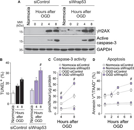
Fig. 2. WRAP53 knockdown enhanced DNA DSBs and neuronal susceptibility to OGD-induced apoptosis.

WRAP53 knockdown was performed by siRNA (siWrap53) transfection for 48 hours, and then neurons were subjected to OGD, as indicated in Fig. 1. (A) Time course of γH2AX and active caspase-3 expression levels detected by Western blotting. GAPDH was probed as loading control. Representative blots are shown. Protein abundance quantification from three different neuronal cultures is shown in fig. S2 (C and D). (B) The percentage of TUNEL-positive siControl and siWrap53-transfected cells was detected by flow cytometry at different time points after OGD. Data are means ± SEM from three different neuronal cultures (#P < 0.05 versus siControl OGD; two-way ANOVA followed by the Bonferroni post hoc test). (C and D) Analysis of caspase-3 activity and neuronal apoptosis in siControl and siWrap53-transfected neurons performed at different time periods after OGD. Data are means ± SEM from three different neuronal cultures (#P < 0.05 versus siControl OGD; two-way ANOVA followed by the Bonferroni post hoc test).
