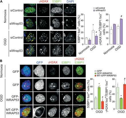
Fig. 4. Nuclear accumulation of WRAP53 promoted DSB repair.

(A) WRAP53 knockdown was performed by siRNA (siWrap53) transfection for 48 hours and neurons were subjected to OGD, as indicated in Fig. 1. Neurons were immunostained for γH2AX and 53BP1 at 8 hours after OGD. Representative images are shown. DAPI, nuclear staining. Scale bar, 5 μm. Quantification of 53BP1 foci–positive neurons (percentage of neurons costaining γH2AX and 53BP1 foci) and percentage of γH2AX foci–positive and 53BP1 foci–negative neurons. The error bars are standard error deviation of 300 measurements from three different neuronal cultures (90 to 110 neurons per culture. *P < 0.05 versus normoxia; #P < 0.05 versus siControl OGD; two-way ANOVA followed by the Bonferroni post hoc test). (B) Neurons were transfected with plasmids encoding GFP, GFP-WRAP53, and MT-GFP-WRAP53 (MT sequence) for 24 hours and were subjected to OGD, as indicated in Fig. 1. Neurons were immunostained for GFP, γH2AX, and 53BP1 at 8 hours after OGD. Representative images are shown. Scale bar, 5 μm. Quantification of 53BP1 foci–positive neurons (percentage of neurons costaining γH2AX and 53BP1 foci) and percentage of γH2AX foci–positive and 53BP1 foci–negative neurons. The error bars are standard error deviation of 30 to 35 measurements from three different neuronal cultures (10 to 15 GFP+ neurons per culture. *P < 0.05 versus GFP; #P < 0.05 versus GFP-WRAP53; one-way ANOVA followed by the Bonferroni post hoc test).
