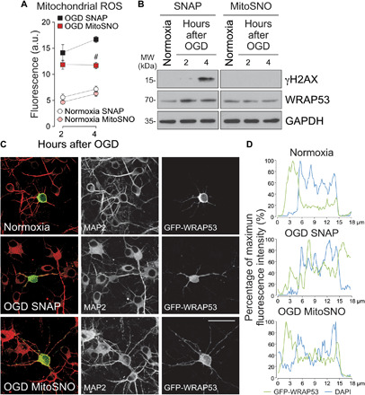
Fig. 6. Mitochondrial generation of ROS triggers WRAP53 nuclear accumulation after OGD.

Neurons were subjected to OGD in the presence of the mitochondrial ROS production inhibitor, MitoSNO (mitochondria-selective S-nitrosating agent). As control, neurons were incubated with SNAP, which do not affect mitochondrial S-nitrosation and respiration. (A) Mitochondrial ROS generation after OGD. The error bars are standard error deviation from three different neuronal cultures (#P < 0.05 versus SNAP OGD; two-way ANOVA followed by the Bonferroni post hoc test). (B) Time course of WRAP53 and γH2AX expression levels as detected by Western blotting. GAPDH was probed as loading control. Representative blots are shown. Protein abundance quantification from three different neuronal cultures is shown in fig. S5D. (C and D) Neurons were transfected with GFP-WRAP53 for 24 hours and were treated with equal doses of SNAP or MitoSNO during OGD (3 hours), as indicated above. (C) Representative images of cortical neurons stained with GFP and MAP2 (neuronal marker). Scale bar, 25 μm. (D) Representative cross-sectional intensity profiles for GFP (green) and DAPI (blue) staining of representative GFP-WRAP53-transfected neurons. a.u., arbitrary units.
