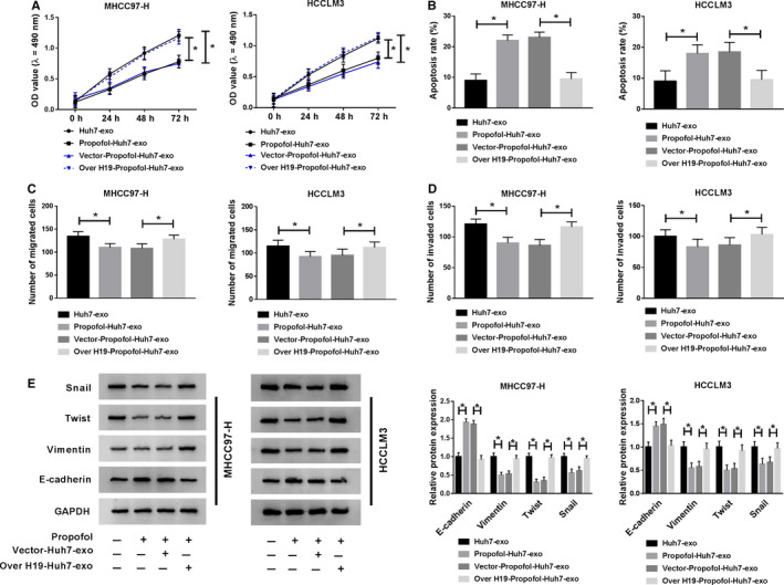
Figure 4.

Exosomal H19 from Huh7 cells enhances the malignant potential of HCC cells. Huh7 cells were treated with Propofol, Vector + Propofol or Over H19 + Propofol, and exosomes were fractionated from the above cells and untreated Huh7 cells. MHCC97‐H and HCCLM3 cells were incubated with exosomes for 24 h. (A) The proliferation of MHCC97‐H and HCCLM3 cells was determined through performing MTT assay. (B) Flow cytometry was applied to detect the apoptosis of MHCC97‐H and HCCLM3 cells. (C and D) The numbers of migrated and invaded HCC cells were counted through transwell assays. (E) Metastasis‐associated markers were measured in HCC cells by Western blot assay. *P < .05
