Figure 6.

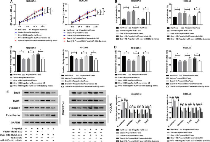
Exosomal H19 from Huh7 cells promotes the proliferation and metastasis and impedes the apoptosis of HCC cells via sponging miR‐520a‐3p. MHCC97‐H and HCCLM3 cells were treated with Huh7‐exo, Propofol‐Huh7‐exo, Vector‐Propofol‐Huh7‐exo, Over H19‐Propofol‐Huh7‐exo, Over H19‐Propofol‐Huh7‐exo + mimic NC or Over H19‐Propofol‐Huh7‐exo + miR‐520a‐3p mimic. (A) The proliferation of HCC cells was determined through MTT assay. (B) The apoptosis of HCC cells was analyzed by flow cytometry. (C and D) The abilities of migration and invasion of HCC cells were measured through conducting transwell assays. (E) Western blot assay was applied to detect the abundance of metastasis‐related proteins in HCC cells. *P < .05
