
FIGURE 5.
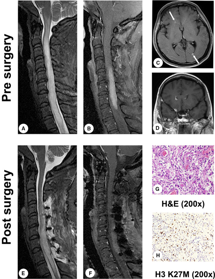
Case 2: Images of an H3 K27M mutation‐positive tumor in a 46‐year‐old female whose main clinical features were dizziness, nausea, vomiting, numbness of the limbs, and weakness of the lower limbs. The histopathology identified an anaplastic astrocytoma. (A, B) Presurgery T2‐weighted and enhanced T1‐weighted magnetic resonance images (MRI). (C, D) Presurgery enhanced T1‐weighted MRI of the brain. (E, F) Postsurgery T2‐weighted and enhanced T1‐weighted MRI. (G) Anaplastic astrocytoma (H&E, 200×). (H) H3 K27M mutation‐positive tumor (H3 K27M, 200×). H&E, hematoxylin and eosin staining