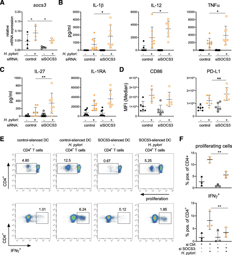
Fig. 6.
SOCS3 silencing shapes H. pylori-induced DC activation and promotes proliferation of T cells. Immature day-7 DCs were re-plated and transfected with siRNA targeting SOCS3 or non-targeting control oligo (both 100 pmol). 48 h post-transfection, H. pylori strain P12 was harvested in PBS and added to the cells at MOI = 5. a Silencing efficiency was analyzed by qRT-PCR after 2 h of infection. One experiment comprising four individual donors is shown. (N = 4) b/c Cytokine secretion of three experiments comprising eight donors (N = 8) was analyzed 24 h post-infection. d Surface marker expression of two experiments comprising six donors (N = 6) was analyzed 24 h after bacterial infection. e/f Control (si Ctrl) or SOCS3-silenced (si SOCS3) DCs were infected with H. pylori (MOI = 5) for 24 h before P/S was added for 6 h. Total CD4+ T cells were magnetically isolated from PBMCs and cultivated with DCs at a ratio of 1:10 (DC:T cell) for 6 days. On day 6, cells were restimulated with PMA/Ionomycin for 4 h and analyzed by means of flow cytometry. Dots represent individual donors (N = 3); mean ± SD is shown. For statistical analysis, one-way ANOVA with Tukey’s post-hoc test was performed. *p < 0.05, **p < 0.01

