Fig. 4.

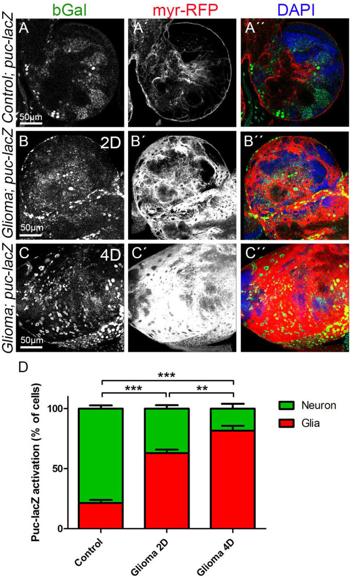
JNK signalling pathway activation in GB. Larval brain sections from third-instar larvae displayed at the same scale. Glial cell bodies and membranes are labelled with UAS-myr-RFP (gray or red in the merge) driven by repo-Gal4 (A–C) JNK signalling pathway reporter puc-lacZ (stained with anti-bGal, gray or green in the merge) in (A) control, (B) glioma induced for 2 days (2D) and (C) glioma induced for 4 days (4D). (A) puc-lacZ reporter signal revealed by bGal staining shows activation of the JNK pathway throughout the brain. 22% of the bGal signal localised in glial cells and most of the signal (78%) localised in the surrounding tissue in control brains. (B,C) puc-lacZ reporter bGal signal shows a progressive activation in GB cells, from 63% after 2 days of GB induction (B) to 80% after 4 days of GB induction (C). (D) Quantification of the % of cells with puc-lacZ activation in glial cells and in the surrounding tissue. Nuclei are marked with DAPI (blue). n=2 independent experiments, n=8 samples analysed for each genotype per experiment. One-way ANOVA with Bonferroni post-hoc test. Error bars show mean±s.d.; **P<0.001, ***P<0.0001. Scale bar size is indicated in all figures. The expression system was active, and the GB induced, during the whole development including both embryonic and larval stages in panel A. In panels B and C animals were raised at 17°C, shifted to 29°C for 2 or 4 days at ∼6 or 2 days after egg laying (AEL), respectively. Genotypes: (A) Gal80ts/repo-Gal4, myr-RFP/puc-lacZ; (B,C) UAS-dEGFRλ, UAS-dp110CAAX; Gal80ts; repo-Gal4, myr-RFP/puc-lacZ.
