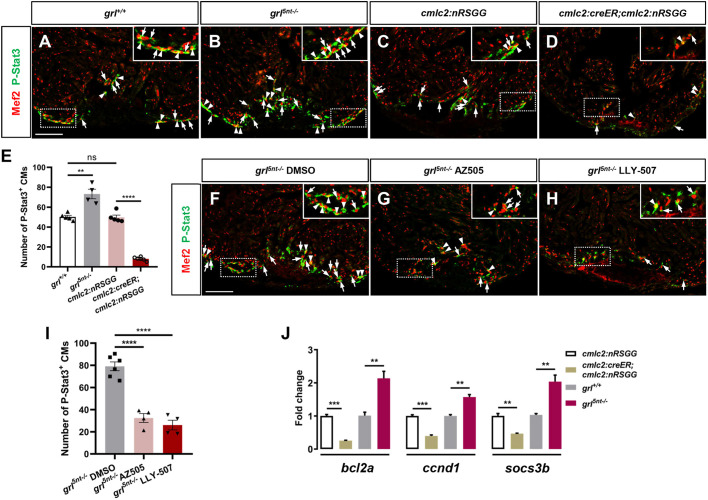
Fig. 7.
Grl-Smyd2 mediates Stat3 activation during heart regeneration. (A-D) Immunofluorescent images of injured ventricles stained with anti-P-Stat3 antibody (green) and anti-Mef2 antibody (red) from WT siblings (A), grl5nt−/− mutant fish (B), 4-HT-treated Tg(cmlc2:nRSGG) control animals (C) and 4-HT-treated Tg(cmlc2:creER;cmlc2:nRGG) animals (D) at 7 dpa. Insets show higher-magnification images of the dashed boxes. Arrowheads indicate P-Stat3 overlapping with Mef2 in CM nuclei; Arrows indicate perinuclear cytosolic P-Stat3 around Mef2 nuclei. P-Stat3 immunostaining was also detectable in non-CM cells in the injured area. (E) Quantification of number of P-Stat3-positive CMs in the injury border zone of 7 dpa ventricles from WT siblings (n=5), grl5nt−/− mutants (n=4), Tg(cmlc2:nRSGG) control fish (n=5), or Tg(cmlc2:creER;cmlc2:nRGG) fish (n=6). (F-H) Immunofluorescent section images of injured ventricles stained with anti-P-Stat3 antibody (green) and anti-Mef2 antibody (red) from DMSO-treated (F), AZ505-treated (G) and LLY-507-treated grl5nt−/− mutant fish (H) at 7 dpa. Insets show higher-magnification images of the dashed boxes. Arrowheads indicate P-Stat3 overlapping with Mef2; Arrows indicate perinuclear cytosolic P-Stat3 around Mef2 nuclei. (I) Quantification of number of P-Stat3-positive CMs in injury border zones at 7 dpa from DMSO-treated (n=6), AZ505-treated (n=4) and LLY-507-treated grl5nt−/− mutant fish (n=4). (J) qPCR analyses of bcl2a, ccnd1 and socs3b in injured hearts extracted from Tg(cmlc2:creER;cmlc2:nRSGG) and Tg(cmlc2:nRSGG) control fish, as well as WT sibling and grl5nt−/− mutant fish (n=3). Data presents as mean±s.e.m. **P<0.01, ***P<0.001, ****P<0.0001, Student's t-test (unpaired, two-tailed). Scale bars: 100 µm.

