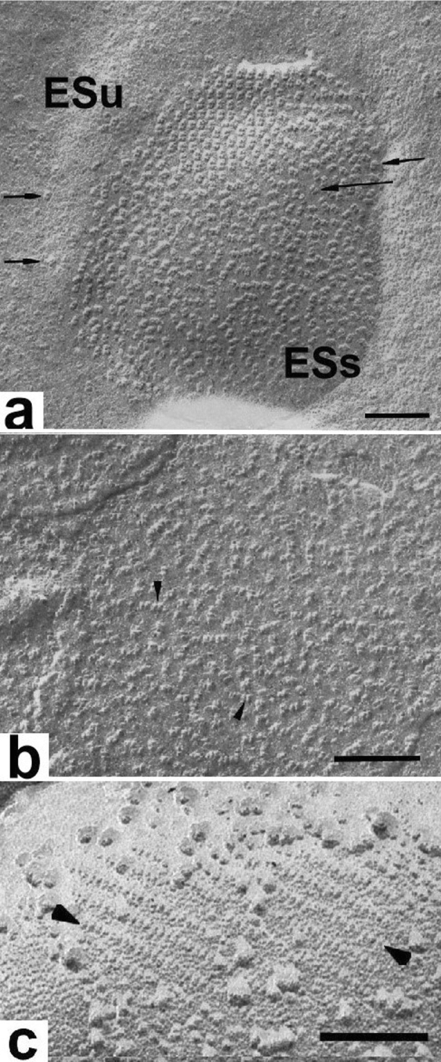
Fig. 16.

a Luminal surface view of a spinach thylakoid exposed by freeze-etching. A central, dimeric particle-rich grana domain (ESs) is surrounded by a stroma thylakoid domain (ESu) with few dimeric particles (arrows). The dimeric particles represent the protruding parts of the oxygen-evolving complexes of the dimeric PSII/LHCII complexes. Some of the dimeric particles form a small lattice. b The dimeric particles (arrows) seen on the luminal surface of a grana membrane after removal of the oxygen-evolving proteins correspond to the core PSII/LHCII complexes, but are smaller than those seen in (a). c Stroma surface of an experimentally unstacked grana thylakoid with arrayed PSII particles (arrows). Together b and c demonstrate that the PSII complexes are integral membrane complexes that extend across the bilayer and protrude from both sides of the bilayer membranes. a From Staehelin (1976), b from Seibert et al. (1987), c from Miller (1976). a Bars 0.1 mm, b 0.1 mm, c 0.2 mm
