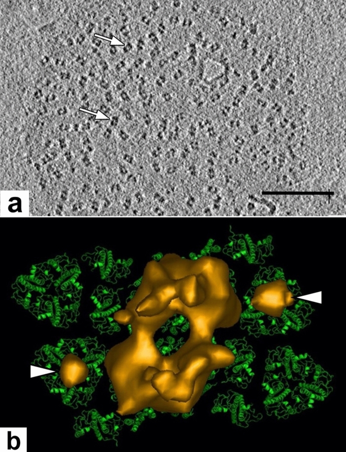
Fig. 18.
Cryo-ET slice image of a grana membrane and an isosurface model of a dimeric PSII complex of spinach. a The dimeric particles (arrows) correspond to protruding parts of PSII/LHCII complexes. b Isosurface model (brown-gold) that shows the extrinsic subunits of the oxygen-evolving complexes of a dimeric PSII complex on the luminal membrane surface superimposed on a pseudo-atomic model of a PSII-LHCII super-complex. The position of the two additional spherical densities (white arrowheads) coincides with the position of the S-type LHCII trimers and probably respresents violaxanthin. From Kouril et al. (2011). a Bar 0.1 mm

