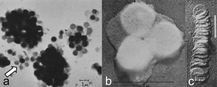
Fig. 2.
Early electron micrographs of isolated chloroplast membranes. a Clusters of grana from individual, disrupted spinach chloroplasts air-dried on grid. The arrow points to a small, separated cluster of grana that appear interconnected by membranes (from Granick and Porter 1947). b Cluster of three, round, gold-shadowed grana “stacks” at high magnification (from Granick and Porter 1947). c Disassembled and gold-shadowed, disrupted granum of the shade plant Aspidistra that resembles a toppled stack of coins. From Steinmann (1952). Bar 1.0 mm

