Fig. 2.
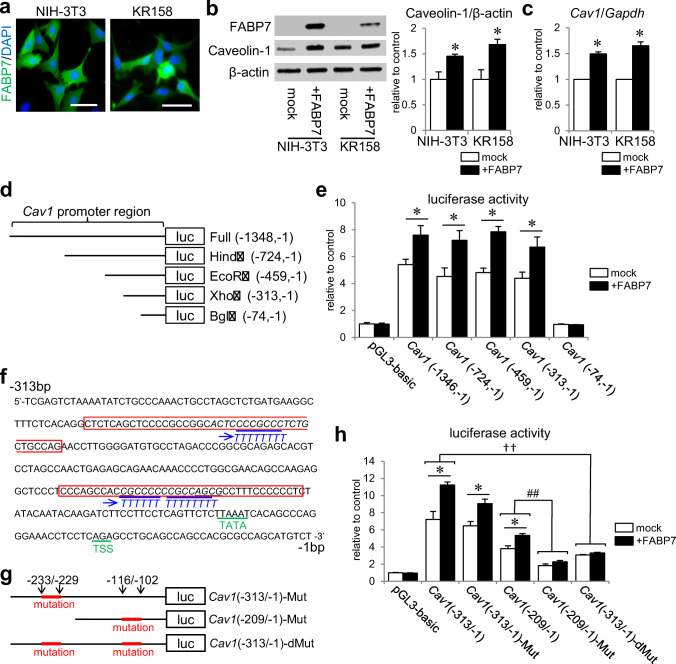
FABP7 affects caveolin-1 promoter activity to regulate caveolin-1 expression. a Immunofluorescence staining of FABP7 (green), and DAPI (blue) in NIH-3T3 cells and KR158 cells. Scale bar: 50 μm. b Western blot for FABP7 and caveolin-1 protein expression in NIH-3T3 cells and KR158 cells. Bar graph shows band density analyzed using NIH-Image J. c qPCR analysis for mRNA expression of Cav1, Lpl, Scpep1, Cav2, and Egfr in NIH-3T3 cells transfected with mock, FABP7, FABP7-NLS (N terminus), and FABP7-NES (N terminus). d Schematic representation of luciferase reporter vectors containing full-length Cav1 promoter, and 5′ deletion mutants using different restriction enzymes. e Luciferase activity assay in NIH-3T3 cell with or without FABP7 overexpression, co-transfected with different reporter vectors of Cav1 promoter. Activity was calculated relative to cells transfected with pGL3-basic luciferase vector. f DNA sequence of Cav1 promoter between – 313-bp and – 1-bp upstream of start codon showing the CG-rich regions. g Schematic representation of luciferase reporter vectors containing mutated Cav1 (− 313/− 1), mutated Cav-1 (− 209/− 1), and double mutations in Cav1 (− 313/− 1). h Luciferase activity assay in NIH3T3 cell with or without FABP7 expression, co-transfected with indicated Cav1 luciferase vectors. Activity was calculated relative to cells transfected with pGL3-basic luciferase vector. Data shown are the means ± s.e.m. and representative of 3 independent experiments. *p < 0.05 versus mock. For panel h, * < 0.05 between mock and NIH-3T3, †† < 0.01, between Cav1 (− 313/− 1) and Cav1 (− 313/− 1) double mutation in both mock and NIH-3T3 with FABP7, ## < 0.01 between Cav1 (− 209/− 1) and Cav1 (− 209/− 1) mutation in both mock and NIH-3T3 with FABP7

