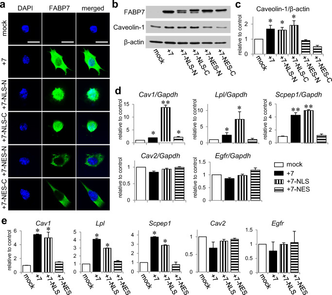
Fig. 4.
Nuclear localization of FABP7 increases caveolin-1 expression via modification of histone acetylation. a Immunofluorescence staining of FABP7 (green), and DAPI (blue) in NIH-3T3 cells transfected with mock, FABP7, FABP7-NLS (in C or N terminus), and FABP7-NES (in C or N terminus). Scale bar: 50 μm. b, c Western blot for FABP7 and caveolin-1 protein expression in NIH-3T3 cells transfected with mock, FABP7, FABP7-NLS (in C or N terminus), and FABP7-NES (in C or N terminus). Bar graph (c) shows band density analyzed using NIH-Image J. d qPCR analysis for mRNA expression of Cav1, Lpl, Scpep1, Cav2, and Egfr in NIH-3T3 cells transfected with mock, FABP7, FABP7-NLS (N terminus), and FABP7-NES (N terminus). e ChIP assays and subsequent qPCR with proximal-1 primer set of Cav1, Lpl, Scpep1, Cav2, and Egfr for the levels of H3K27ac in NIH3T3 cells transfected with mock, FABP7, FABP7-NLS (N terminus), and FABP7-NES (N terminus). Data shown are the means ± s.e.m. and representative of 3 independent experiments. *p < 0.05, **p < 0.01 versus mock

