Fig. 7.
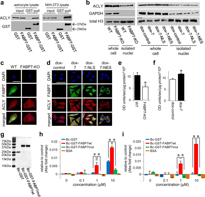
FABP7 interacts and regulates ATP-citrate lyase. a GST pull-down analysis and western blot to confirm the interaction of FABP7 and ACLY using FABP7-KO primary cultured astrocyte and NIH-3T3 cell lysate. b Western blot for ACLY expression in whole cell or isolated nuclei of WT and FABP7-KO primary cultured astrocytes or of NIH-3T3 cells with doxycycline-induced control, FABP7, FABP7-NLS, and FABP7-NES. c, d Immunofluorescence staining of FABP7 (green), ACLY (red), and DAPI (blue) in WT and FABP7-KO primary cultured astrocytes (c) or in NIH-3T3 cells with doxycycline-induced control, FABP7, FABP7-NLS, and FABP7-NES (d). Scale bar: 50 μm. e, f Measurement for ACLY activity in WT and FABP7-KO primary cultured astrocytes (e) or in NIH-3T3 cells with doxycycline-induced control and FABP7 (f). The levels were normalized by the protein concentration. g Western blot using GST antibody to confirm recombinant protein. h, i Measurement for ACLY activity with recombinant protein and BSA using FABP7-KO primary cultured astrocytes lysate (h) or NIH-3T3 cell lysate (i). Graph shows the difference compared to non-treated. Raw data is shown in Fig. S4e and S4f. Data shown are the means ± s.e.m. and representative of 3 independent experiments. *p < 0.05 versus WT or control. For Fig. 7h and i, analysis was performed in the group of between GST-treated and FABP7wt treated or between FABPwt-treated and FABP7mut-treated

