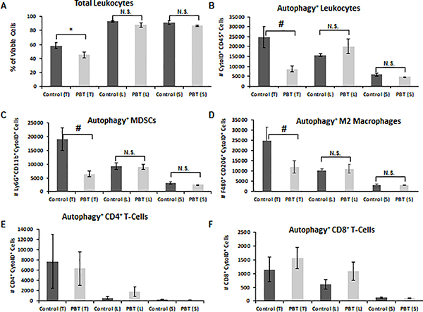
Figure 2. DFMO and Trimer PTI reduced autophagy in immunosuppressive leukocytes.
Mice were orthotopically injected in the mammary fat pad with 2 × 10⁴ 4T1 mammary carcinoma cells. Three weeks following injection of 4T1 cells, treatment was initiated with either control vehicle or PBT. Mice were sacrificed after three weeks of treatment, and equal numbers of cells from tumors (T), lungs (L), and spleens (S) were analyzed by flow cytometry for, A: total leukocytes (CD45+), B: autophagic activity in CD45+ leukocytes (CytoID+CD45+), C: autophagy+ MDSCs (CytoID+Gr1+CD11b+), D: autophagy+ M2 macrophages (CytoID+F480+CD206+), E; autophagy+ CD4+ T-cells (CytoID+CD4+), F: autophagy+ CD8+ T-cells (CytoID+CD8+). n = 10 per group ± SEM; * = p ≤ 0.05 and # = p ≤ 0.01 compared to vehicle treated mice.

