Figure 2.
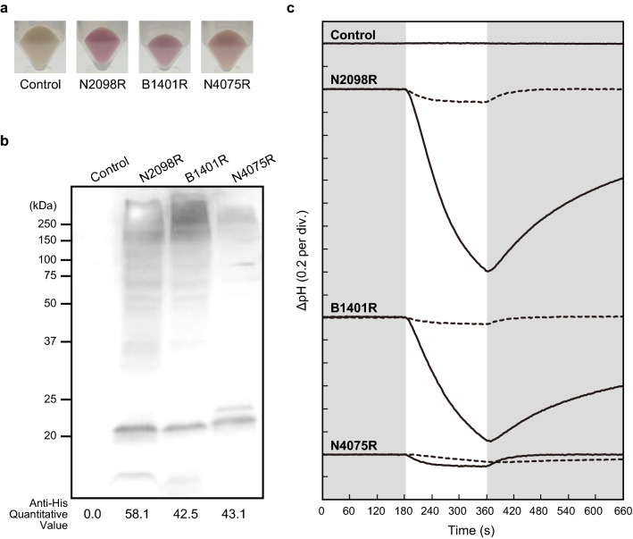
Light-induced changes of the pH of suspensions of Escherichia coli expressing a rhodopsin (N2098R, B1401R, and N4075R) from the novel CyR clade. (a) The pellet color of CyRs. (b) Detection of protein expression of CyRs by western blots using an anti-His-tag antibody. These proteins were expressed in E. coli cells with a His-tag at the C-terminal. The monomer-band of CyRs (around 22 kDa) were quantified using ImageJ software. (c) The changes in pH in the absence (solid line) and presence (broken line) of CCCP are shown. The numbers in parentheses are the pH units of y-axis divisions. All measurements were performed under the dark condition (gray shading) with illumination at 520 ± 10 nm for 3 min (white shading). E. coli cells containing the pET21a plasmid vector alone were simultaneously analyzed as a negative control.

