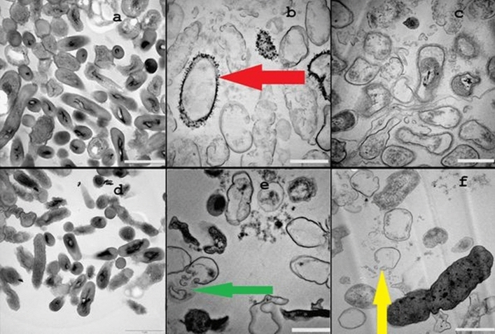
Figure 6.
Representative TEM micrographs of untreated B. subtilis CN2 cells (a), showing intact and high electron density morphology, 100 µg/mL (b), 125 µg/mL (c) dosage of Cu2O NPs treated cells indicating cytoplasmic injury with disintegrated outer membrane (f, yellow arrow). P. aeruginosa CB1 cells treated with 100 µg/mL (e), 125 µg/mL (f) dosage of Cu2O NPs and untreated control (d). Considerable size of adhered nanoparticles was observed (b, red arrow) attached to the surface of the cells of the bacteria, and disrupted cell wall and membrane leakage was observed (e, green arrow). Scale bar is 1 µm.

