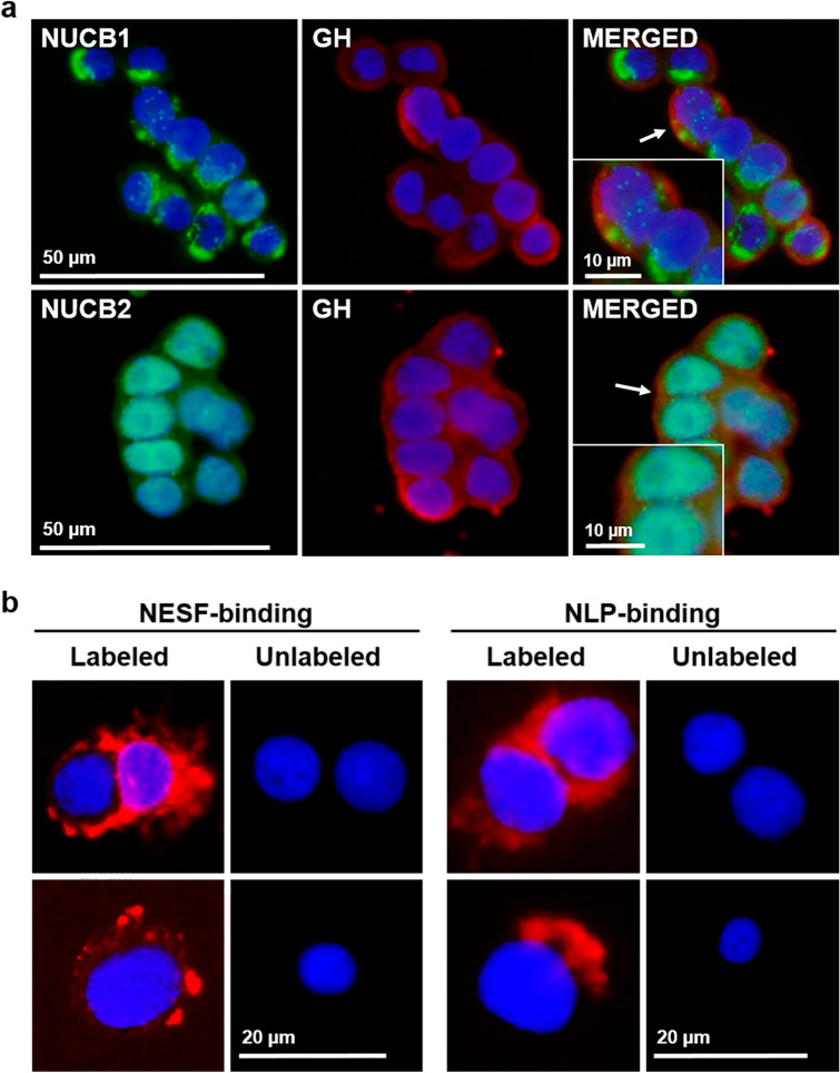
Figure 2.
(a) NUCB1/NLP and NUCB2/NESF colocalizes with GH in somatotrophs. Representative images of immunofluorescence detection of NUCB1 (green), NUCB2 (green) and GH (red) in GH3 cells. Cells were counterstained with DAPI (blue) and the images were acquired at 40X magnification. Representative and magnified images corresponding to the areas indicated with an arrow in the merged figure are shown in the inset. (b) NESF and NLP bind to mammalian somatotroph cells. Representative images of NESF-binding (left) and NLP-binding (right) detection (in red) in the surface of GH3 cells after incubation for 1 h with 1 nM CF568-labeled NESF or NLP, or unlabeled-peptides, followed by several washes with PBS. GH3 cells were counterstained with DAPI (in blue), and images were acquired at 40X magnification.

