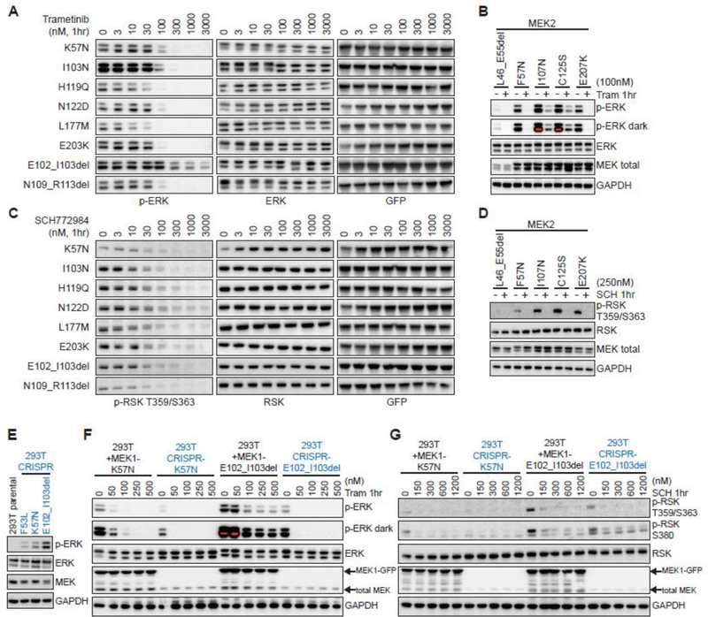
Figure 5. MEK1/2 mutants are variably sensitive to MEK and ERK inhibition.
A-B, Hotspot and activating MEK1 (A) or MEK2 (B) missense and in-frame deletion mutants were expressed in 293H cells for 24hr, then treated for 1hr with vehicle or (A) the MEK inhibitor trametinib (33000nM) or (B) trametinib at 100nM followed by assessment of p-ERK expression by western blot. C-D, Hotspot and activating MEK1 (C) or MEK2 (D) missense and in-frame deletion mutants were expressed in 293H cells for 24hr, then treated for 1hr with vehicle or (C) the ERK inhibitor SCH772984 (3–3000nM) or (D) SCH772984 at 250nM followed by assessment of p-RSK expression by western blot. E, p-ERK expression in 293T cells in which MEK1 F53L, K57N or MEK E102_I103del mutants were knocked-in using CRISPR/Cas9. F-G, MEK1 K57N or E102_103del constructs were overexpressed in 293T and compared to their respective 293T MEK1 CRISPR knock-in derived cell lines (in blue) for changes in p-ERK (F) or p-RSK (G) at baseline and after 1hr treatment with vehicle or increasing doses of (F) trametinib (50–500nM) or (G) SCH772984 (150–1200nM).

