
Figure 2.
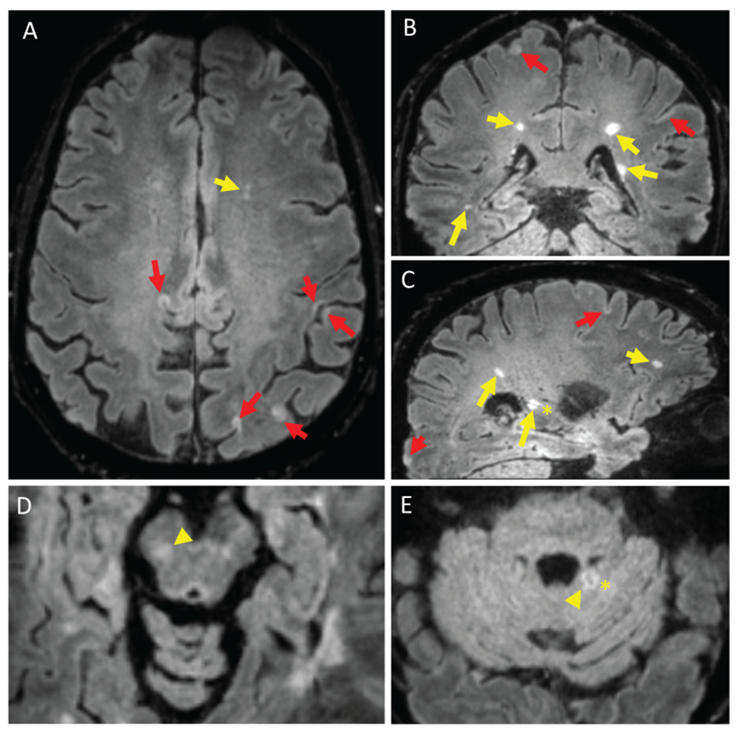
Inversion recovery susceptibility weighted imaging with enhanced T2 weighting (IR-SWIET) images. IR-SWIET is a 3D sequence reconstructed with isotropic voxels, allowing for reconstruction in axial (A), coronal (B), and sagittal (C) planes. IR-SWIET images have suppressed CSF signal, making cortical lesions easier to see. (A-C) White matter lesions (yellow arrows) and cortical lesions (red arrows) are conspicuous and central veins (asterisk) can be seen in some lesions. Lesions in the brainstem (D, yellow arrowhead) and cerebellum (E, yellow arrowhead) are also observed. All images are medians of four IR-SWIET acquisitions.