
Figure 3.
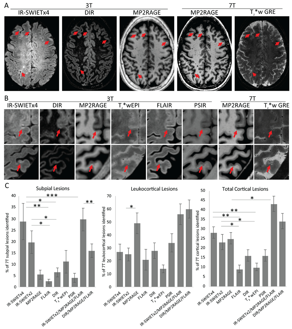
IR-SWIET improves subpial lesion detection. A) Subpial lesions (red arrow) are well seen on IR-SWIET images compared to other 3T images and are confirmed as lesions on 7T images. B) High magnification view of two subpial lesions identified on IR-SWIET but which are more subtle on other 3T images and are confirmed on 7T images. C) Quantification of sensitivity of individual 3T sequences and 3T multicontrast reads compared to lesions identified on 7T images. *=p<0.05, **=p<0.01, ***=p<0.001, IR-SWIET – inversion recovery susceptibility weighted imaging with enhanced T2 weighting (x2 - median of two acquisitions, x4 – median of four acquisitions), DIR – double inversion recovery, MP2RAGE – magnetization-prepared 2 rapid gradient echo, T2*wEPI – T2* weighted segmented echo-planar imaging, FLAIR – fluid-attenuated inversion recovery, PSIR – phase-sensitive inversion recovery, GRE – gradient recalled echo.