
Figure 1.
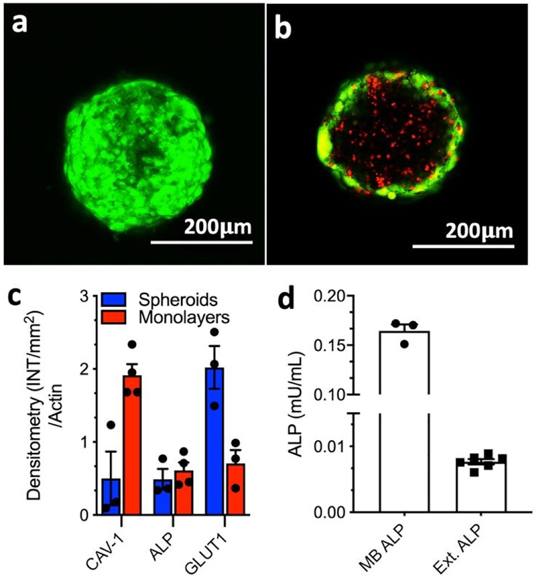
Characterization of the HS587T spheroids: Representative confocal microscopy images of (a) spheroid surface (whole spheroid projection) and (b) core (single focal plane/z-stack) stained for live (green, calcein AM) and dead (red, propidium iodide) cells; (c) Densitometry quantification of Western-Blot analysis of ALP, CAV1, and GLUT1 expression by the 3D spheroids and 2D cell culture, normalized to the total β-actin protein content; (d) expression of membrane-bound (MB) and extracellular (Ext.) ALP in spheroids.