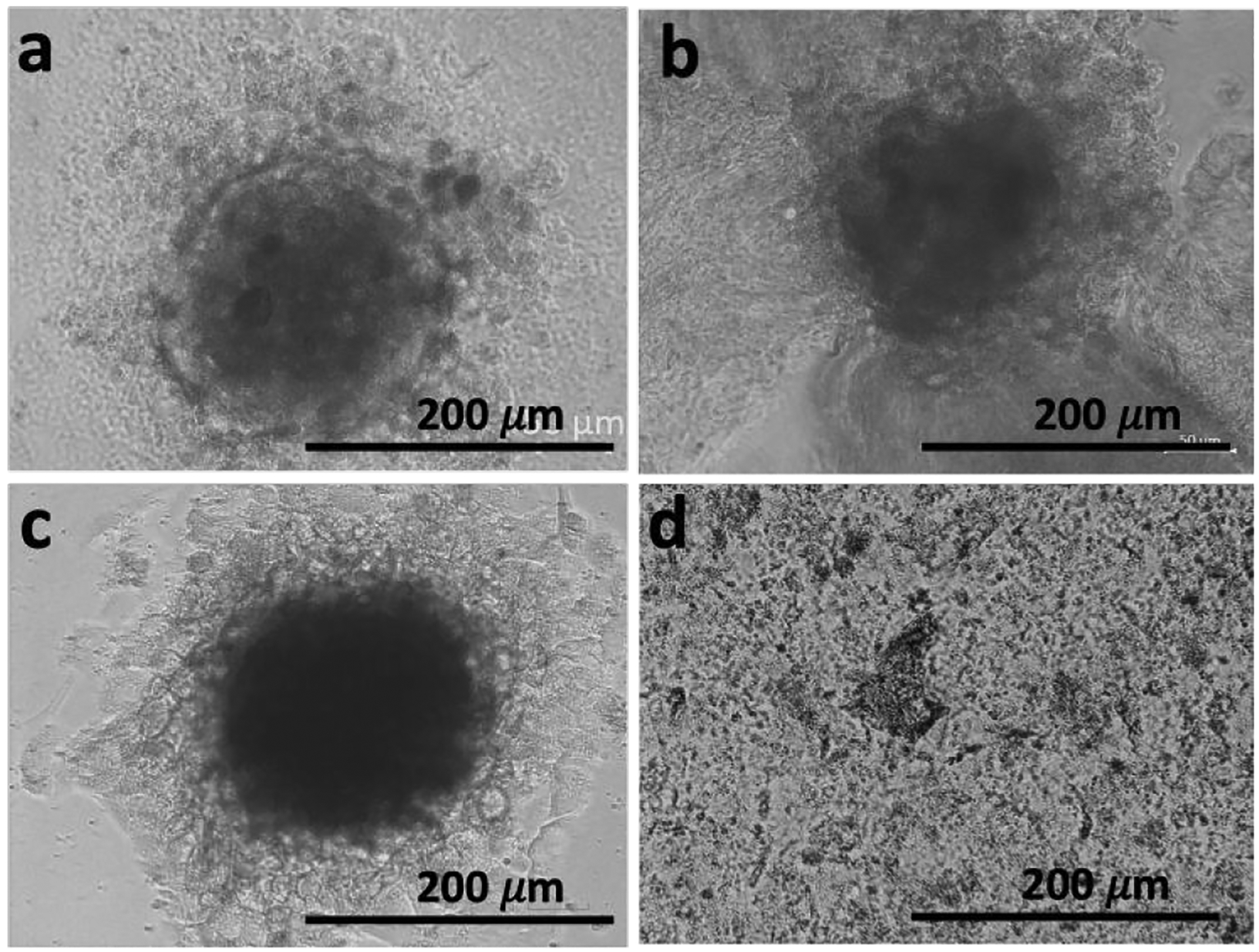
Figure 4.

Spheroid relapse shown by replating the spheroids: representative transmitted-light microscope images of (a) untreated spheroid (control) and (b) spheroid exposed to 1 (1 mM, 48 h) that were re-plated (c control and d treated) in adherent well plates for 48 h.
